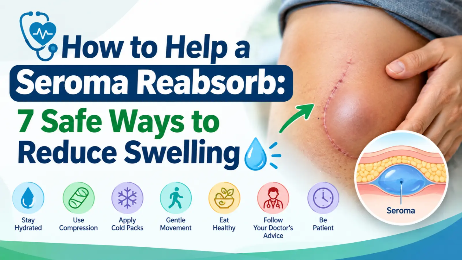
A seroma is that soft, fluid-filled swelling that shows up near a surgical site days or weeks after your procedure can be alarming if you don’t know what it is.
The good news: seromas are one of the most common post-surgical complications, and the vast majority resolve on their own without any dramatic intervention.
If you’ve recently had surgery (breast surgery, liposuction, a tummy tuck, hernia repair, or another procedure involving significant tissue disruption) and you’re now watching a pocket of fluid slowly shrink or wondering why it isn’t shrinking fast enough this guide walks through exactly what helps a seroma reabsorb.
We’ll cover the at-home strategies that actually make a difference, the medical treatments your doctor might recommend, realistic timelines for recovery, and the warning signs that mean it’s time to stop waiting and call your provider.
Understanding Seromas
What Exactly Is a Seroma?
A seroma is a pocket of clear or straw-colored fluid that collects under the skin, typically in a space left behind after tissue has been surgically removed, lifted, or separated from underlying structures.
The fluid itself is primarily serum, the watery component of blood plasma mixed with lymphatic fluid, rather than blood or pus.
How a Seroma Forms
Surgery disrupts small blood and lymphatic vessels as tissue is cut, lifted, or repositioned.
In the days that follow, the body’s healing response sends fluid to the site, but when there’s empty space (often called “dead space”) left behind and the lymphatic vessels haven’t yet reconnected or healed that fluid has nowhere to drain.
Instead, it pools and collects, forming a seroma.
Seroma vs. Other Post-Surgical Swelling
It’s easy to confuse a seroma with other complications, but they behave differently:
- Seroma vs. hematoma: A hematoma is a collection of blood, not clear fluid; it often appears more red/purple, more painful, and forms faster (within hours, not days).
- Seroma vs. abscess: An abscess involves infection and pus, and typically comes with redness, warmth, and fever a plain seroma should not.
- Seroma vs. general edema: Edema is more diffuse, whole-area swelling, while a seroma is usually a distinct, localized, fluid-filled pocket you can often feel move under the skin.
Common Causes and Procedures
Seromas are most associated with surgeries that involve large areas of tissue separation, including:
- Mastectomy and lumpectomy
- Lymph node removal/dissection
- Liposuction
- Abdominoplasty (tummy tuck)
- Hernia repair
- Large excisions or flap surgeries
- Occasionally, blunt trauma or injury without surgery (this is well documented in veterinary medicine too, particularly in horses and dogs)
Typical Timeline of Formation
Most seromas appear 7 to 10 days after surgery often right around the time surgical drains are removed.
This timing isn’t a coincidence: drains are placed specifically to prevent fluid buildup, so once they come out, any remaining fluid production has nowhere to go but into that dead space.
Signs and Symptoms
Recognizing a seroma and distinguishing it from something more serious starts with knowing what to look for.
The most common signs include:
- A soft, balloon-like swelling near the surgical site, usually appearing smooth and well-defined under the skin
- A sensation of fluid movement, sometimes described as feeling like a water balloon or a squishy pocket that shifts slightly when pressed
- Clear or straw-colored fluid, if any is visibly draining from the incision
- Mild discomfort, tightness, or pressure, particularly as the seroma grows larger
- Localized swelling that’s more defined than the general puffiness typical of normal post-op healing
Size can vary significantly.
Small seromas may be barely noticeable, roughly the size of a grape, while larger ones can grow to the size of a golf ball, an orange, or even bigger in extensive procedures like tummy tucks or mastectomies.
What a normal seroma should NOT include:
- Redness or warmth spreading across the skin
- Fever or chills
- Sharp or worsening pain (mild pressure is normal; escalating pain is not)
- Cloudy, yellow, or foul-smelling drainage
- Rapid growth over just a few hours
If your swelling is soft, painless or only mildly uncomfortable, and the surrounding skin looks normal, you’re likely dealing with a straightforward seroma.
Any deviation from that, especially signs pointing toward infection, is a signal to contact your surgeon rather than waiting it out.
Diagnosis
While many seromas are recognizable through symptoms alone, proper diagnosis helps rule out complications and confirms the right course of action.
Physical Examination
In most cases, your surgeon or healthcare provider can diagnose a seroma simply by examining the area.
They’ll typically check for the characteristic soft, fluid-filled swelling, note whether it moves or shifts under gentle pressure, and assess the surrounding skin for signs of infection like redness or warmth.
They’ll also ask about your timeline when the swelling appeared relative to your surgery date since seromas follow a fairly predictable pattern.
When Imaging Is Used
For larger, deeper, or ambiguous cases, your provider may order an ultrasound.
This is the most common imaging tool used to confirm a seroma, as it can clearly show a fluid-filled pocket versus a solid mass, help estimate the volume of fluid present, and rule out other possibilities like a hematoma, abscess, or, more rarely, a tumor.
Ultrasound is especially useful when:
- The swelling is large or growing quickly
- There’s uncertainty about whether it’s fluid or solid tissue
- The seroma is in a location where a tumor recurrence needs to be ruled out (such as after breast cancer surgery)
- Your provider is deciding whether aspiration is appropriate
When Imaging Isn’t Necessary
For small, classic-presentation seromas with a clear post-surgical timeline and no red flags, many providers are comfortable diagnosing based on exam alone, without imaging, and simply monitoring for changes over the following weeks.
Non-Invasive Methods to Support Reabsorption
Most seromas respond well to simple, consistent home care. Here’s what actually helps.
Compression is the single most effective tool.
A compression garment, binder, or elastic wrap applied over the area closes the dead space where fluid collects, encouraging reabsorption instead of continued pooling.
Following your surgeon’s guidance on tightness and duration too loose won’t help, too tight can restrict circulation.
Rest and activity modification reduce strain on healing tissue.
Avoid heavy lifting, vigorous exercise, or repetitive motion near the site, as excess movement can stimulate more fluid production.
Elevation helps when the seroma is on a limb.
Raising the area above heart level during rest reduces fluid accumulation, similar to standard swelling management.
Warm compresses, applied gently a few times daily, can encourage circulation and support the body’s natural fluid reabsorption.
Check timing with your provider warmth is usually introduced once initial post-op inflammation has settled, not immediately after surgery.
Gentle massage, only once your surgeon approves it, can help guide fluid toward the lymphatic system.
This should never be attempted on a fresh incision or without professional clearance, as improper technique can irritate the area.
Hydration and nutrition support overall tissue healing and lymphatic function, though they play a supporting role rather than a primary one.
Time remains the most underrated factor.
Small seromas often shrink noticeably within two to four weeks; patience, paired with consistent compression, is often the entire treatment plan.
Medical Interventions
When home care isn’t enough, or a seroma is large, painful, or persistent, your doctor has several options.
Needle aspiration is the most common intervention.
Using a syringe, your provider draws the fluid out directly, which can offer immediate relief from pressure and discomfort.
However, aspiration carries a meaningful drawback: it doesn’t address the underlying dead space, so fluid often reaccumulates, sometimes within days.
Each aspiration also introduces a small infection risk, since it involves puncturing the skin.
Repeated aspirations are sometimes necessary for seromas that keep refilling.
Providers generally try to limit how many times they’ll aspirate the same seroma, both due to infection risk and because repeated drainage can occasionally worsen chronic fluid buildup.
Sclerotherapy is used for persistent or recurrent sarcomas that don’t respond to aspiration alone.
A sclerosing agent is injected into the cavity after drainage, irritating the tissue walls so they stick together and close off the space, preventing further fluid collection.
Surgical drainage or drain placement may be needed for large, deep, or repeatedly recurring seromas.
In some cases, a small drain is reinserted to continuously remove fluid over several days, allowing the tissue to heal closed.
Combined approaches are common in practice: aspiration paired with compression, or drainage followed by a stricter compression protocol, often produces better long-term results than any single method alone.
Your surgeon will weigh seroma size, symptoms, recurrence history, and infection risk when deciding which path makes sense for your situation.
Prevention Strategies (Pre- and Post-Procedure)
While not every seroma is preventable, several strategies significantly reduce the risk, both in the operating room and during recovery.
Surgical drain placement is the most common preventive measure.
Surgeons often place temporary drainage tubes during procedures with a high seroma risk (like mastectomies, tummy tucks, or large flap surgeries) to continuously remove fluid before it has a chance to pool in the dead space left behind.
Quilting sutures are an increasingly used technique, particularly in abdominoplasty and breast surgery.
These internal stitches tack the skin down to the underlying tissue, closing off the dead space directly at the source rather than relying on drains or compression afterward.
Studies have shown this technique can meaningfully reduce seroma rates in certain procedures.
Tissue glue or fibrin sealants are sometimes applied during surgery to help bond tissue layers together, reducing the empty space where fluid could otherwise collect.
Early, consistent post-op compression starts the moment surgical dressings allow it, rather than waiting for a seroma to appear.
Many surgeons build compression garments into standard post-op protocols for higher-risk procedures.
Structured activity restriction in the first one to two weeks avoiding lifting, reaching, or straining helps minimize the tissue movement that can stimulate fluid production.
Patient education matters more than it might seem.
Surgeons who clearly explain compression schedules, activity limits, and warning signs tend to see better compliance, and better compliance is directly linked to lower seroma rates.
Complications to Watch For
Most seromas resolve without incident, but a small number develop complications worth knowing about.
Infection is the most serious risk.
Because a seroma is essentially stagnant fluid sitting under the skin, it creates an environment where bacteria can thrive if introduced, whether through the original incision or during aspiration.
Warning signs include redness, warmth, increasing pain, fever, chills, or cloudy/foul-smelling drainage.
Infected seromas typically require antibiotics and, often, drainage.
Skin breakdown or necrosis can occur when a seroma grows large enough to put sustained pressure on the overlying skin, restricting blood flow to that tissue.
Over time, this pressure can cause the skin to thin, break down, or in severe cases, die, particularly if the seroma is left unaddressed for an extended period.
Chronic or recurrent seromas are ones that keep refilling despite aspiration or don’t resolve within the expected timeframe (typically several months).
These often need a more aggressive approach, such as sclerotherapy or surgical intervention, rather than continued watchful waiting.
Capsule formation happens when a long-standing seroma becomes “encapsulated” ; the body forms a fibrous wall around the fluid pocket, similar to a cyst.
Once encapsulated, a seroma is far less likely to reabsorb on its own and often won’t respond well to simple aspiration, since the capsule tends to refill or require surgical removal entirely.
Masking other issues is a less common but important concern, particularly after cancer-related surgery.
A persistent or unusual swelling should be evaluated to rule out recurrence, rather than assumed to be a straightforward seroma.
Recovery Timeline Expectations
One of the most common questions people have about seromas is simply: how long will this take?
The honest answer is that it varies significantly depending on size, location, and individual healing.
Small seromas, those roughly grape- to golf-ball-sized, often begin shrinking noticeably within one to two weeks and may fully reabsorb within three to four weeks, especially with consistent compression.
Larger seromas, such as those following extensive procedures like tummy tucks, mastectomies, or large flap surgeries, typically take longer.
It’s not unusual for these to take several weeks to a few months to fully resolve, and in some documented cases, up to a year.
Factors that slow reabsorption include:
- Inconsistent or improper use of compression garments
- Continued strenuous activity that irritates the area
- Larger initial fluid volume
- The specific type of surgery performed (procedures with more extensive tissue disruption tend to have longer seroma timelines)
- Individual healing factors, including age, circulation, and overall health
Factors that support faster resolution include:
- Strict adherence to compression protocols
- Early, appropriate activity restriction
- No secondary complications like infection
It’s worth setting realistic expectations early: a seroma that’s still present at six or eight weeks isn’t necessarily abnormal, particularly after major surgery.
Slow, steady shrinkage is a good sign, even if the pace feels frustrating.
What matters most is the trend, not the exact timeline.
When to Contact Your Doctor (Red Flags Recap)
While most seromas are a normal, manageable part of recovery, certain symptoms should never be brushed off or “waited out.”
Knowing when to escalate care can prevent a minor complication from becoming a serious one.
Contact your doctor promptly if you notice:
- Rapid increase in size especially if the swelling grows noticeably within hours rather than gradually over days
- Redness or warmth spreading across the skin around the seroma, which can indicate infection
- Fever or chills, even mild ones, which should never be ignored in the context of a recent surgical site
- Significant or worsening pain mild pressure or tightness is expected, but sharp, escalating, or throbbing pain is not
- Cloudy, yellow, or foul-smelling drainage from the incision, rather than clear or straw-colored fluid
- Skin that appears thin, shiny, or discolored over the seroma, which can signal pressure-related skin breakdown
- No improvement after several weeks, particularly for smaller seromas that should be trending smaller, not staying the same size or growing
A general rule of thumb: if something about the swelling feels different from what your surgical team described as “normal healing,” it’s always reasonable to call and ask, even if it turns out to be nothing.
Surgeons expect and welcome these check-ins, especially in the first few weeks after a procedure.
Trust your instincts here.
You know your body and your recovery better than anyone reading a general guide, and a quick call is far better than a delayed one.
Frequently Asked Questions
Can a seroma go away without treatment?
Yes, most small to moderate seromas resolve entirely on their own, with the body gradually reabsorbing the fluid over several weeks.
Treatment beyond compression and monitoring often isn’t necessary unless the seroma is large, painful, or shows signs of infection.
Is it safe to massage a seroma myself?
Only with your surgeon’s explicit approval.
Massaging too early, too aggressively, or over a fresh incision can irritate tissue or increase fluid production rather than reduce it.
If cleared, gentle, guided massage can help encourage drainage toward the lymphatic system.
Does a seroma always need to be drained?
No.
Many seromas, especially smaller ones, are left alone and simply monitored, since aspiration carries its own risks, including infection and fluid recurrence.
Draining is typically reserved for seromas causing significant discomfort, pressure, or risk of skin complications.
Can seromas come back after aspiration?
Yes, this is fairly common.
Because aspiration removes fluid without closing the underlying dead space, the area can refill, sometimes within days.
This is why some seromas require repeated aspiration or a different approach like sclerotherapy.
How long is too long for a seroma to persist?
There’s no universal cutoff, but if a seroma hasn’t started shrinking after several weeks, or is still present at the two-to-three-month mark without any improvement, it’s worth discussing further options with your doctor rather than continuing to wait it out.
Conclusion
Dealing with a seroma after surgery can feel unsettling, but in most cases, it’s a normal and manageable part of the healing process.
The core strategy is straightforward: consistent compression, sensible activity restriction, elevation when applicable, and patience.
Small seromas often shrink within a few weeks, while larger ones may take longer.
Stay attentive to red flags like infection, rapid growth, or persistent pain, and reach out to your healthcare provider when needed. Most seromas resolve fully with proper care and time.
This article is for general informational purposes and is not a substitute for personalized medical advice from your healthcare provider.